1/3
Export Ready
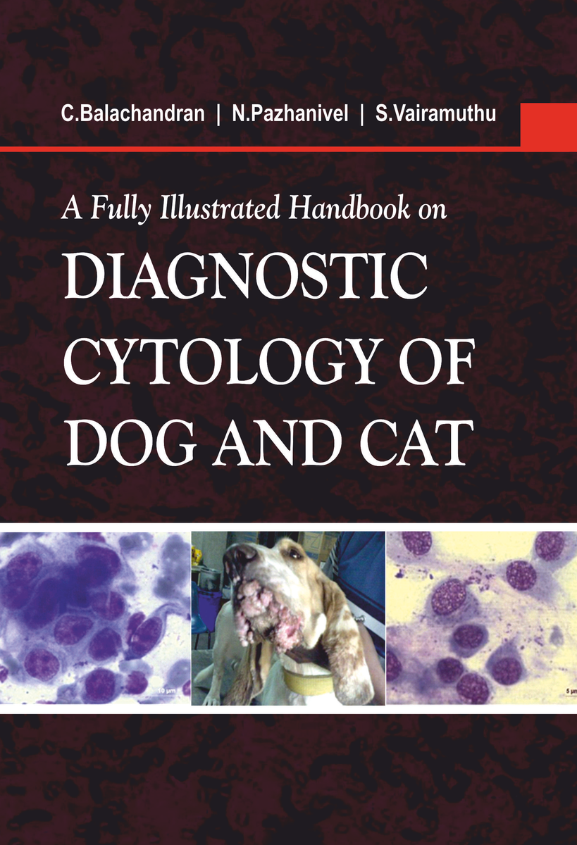
A Fully Illustrated Handbook on Diagnostic Cytology of Dog and Cat (Hardback, C.Balachandran, N.Pazhanivel &.S.Vairamuthu)
Sold by nipa genx electronic resources & solutions · Service Provider , Manufacturer, Professional Services, Brand Owner, Distributor, Exporter, Importer, Wholesaler, Startup · North Delhi, Delhi, India
Price
₹ 198.00
/Unit
In stock
Inclusive of all taxes
Minimum order:
1 Unit
Sample request
Sample price: 0