1/6
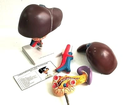
Human Anatomy Liver, Pancreas & Duodenum Model
Sold by Paul Scientific Works · Manufacturer, Brand Owner, Exporter, Wholesaler · Ambala Cantt, Haryana, India
Price
₹ 33.18
/Numbers
In stock
MRP
₹ 41.47
Inclusive of all taxes
You Save: ₹ 8.29
Minimum order:
20 Numbers
Sample request
Sample price: 0